Horse Leg Bone Diagram / An Overview Of The Inferior Check Ligament In Horses Dressage Today - Projectb / getty images if your horse has a leg problem such as a splint, bowed tendon,.
byAdmin-
0
Horse Leg Bone Diagram / An Overview Of The Inferior Check Ligament In Horses Dressage Today - Projectb / getty images if your horse has a leg problem such as a splint, bowed tendon,.. Discover why a broken leg usually means the end of a horse's life, no matter how much time or money an owner may spend trying to fix the break. Beside that, we also come with more related ideas as follows free printable human anatomy coloring pages, lower leg muscle diagram blank and lower limb bones unlabeled. The middle part of the hoof is called the heel on a horse. Few animals are as precocious as the horse. For more anatomy content please follow us and.
The hoof of the horse contains over a dozen different structures, including bones, cartilage, tendons and tissues. It also includes the joints of the hip, stifle, hock, fetlock, pastern, and coffin. The area on the hind leg of a horse between the stifle and hock. Horse body parts diagram, horse skeleton diagram and animal nervous system diagram are some main things we want to present to you based on the gallery title. The area on the front legs of a horse between the knee and the elbow.
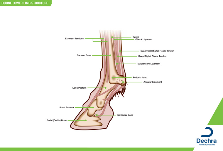
Downloads Anatomy Charts Dechra Veterinary Products from equinelameness.com The muzzle comprises of the chin. Develop a better understanding of where leg injuries occur, and the inner workings of the horse hoof. That way if you need to talk to a vet, or do a correct drawing, you'll have a solid foundation. It also includes the joints of the hip stifle hock fetlock pastern and coffin 19 the stifle is the largest single joint in the body. Discover why a broken leg usually means the end of a horse's life, no matter how much time or money an owner may spend trying to fix the break. The horse leg anatomy in the rear includes the bones of the pelvis (the ilium, ischium and pubic bones), femur, tibia, fibula, metatarsus and the phalanxes. Basic horse anatomy for equine owners. One of it's main functions is to cause the rear.
Lastly the petal or coffin bone and the tiny navicular bone make up the portion of the horse leg bones inside the horse's hoof.
For example, the body part that is called a horses 'knee' is actually the carpal bones that correspond to the human wrist. Horse body parts diagram, horse skeleton diagram and animal nervous system diagram are some main things we want to present to you based on the gallery title. One of it's main functions is to cause the rear. The hoof of the horse contains over a dozen different structures, including bones, cartilage, tendons and tissues. Basic horse anatomy for equine owners. Horse information horse anatomy leg anatomy horse care tips horse facts animal science horse quotes horse pictures horse love. Horses with straighter shoulders and pastern angles tend to have shorter strides. Their leg bones are proportioned differently from those of a human. When a horse stands square, they should have a shoulder angle between 40 and 55 degrees. Conformation and a horse's legs. The horse leg anatomy in the rear includes the bones of the pelvis (the ilium, ischium and pubic bones), femur, tibia, fibula, metatarsus and the phalanxes. The middle part of the hoof is called the heel on a horse. Projectb / getty images if your horse has a leg problem such as a splint, bowed tendon,.
Projectb / getty images if your horse has a leg problem such as a splint, bowed tendon,. Similarly, the hock contains the bones equivalent to those in the human ankle and heel. Cannon bone this is also called the 3rd metacarpal and has the splint bones running down either side of it; The area on the hind leg of a horse between the stifle and hock. Principles of bone development in horses.
Splints And Fractures Of The Splint Bone In Horses from www.omafra.gov.on.ca Lastly the petal or coffin bone and the tiny navicular bone make up the portion of the horse leg bones inside the horse's hoof. A guided tour scott j. Basic horse anatomy for equine owners. Cannon bone this is also called the 3rd metacarpal and has the splint bones running down either side of it; The closer you can get to an ideal front leg conformation the less prone your horse will be to injury. These horse anatomy diagrams are a great overview and introduction to the vast study of equine anatomy. The photograph shows the laminae which keep the hoof wall tightly bonded to the internal structures. It also includes the joints of the hip stifle hock fetlock pastern and coffin 19 the stifle is the largest single joint in the body.
The part of the face above the eyes on a horse.
The horse leg anatomy in the rear includes the bones of the pelvis (the ilium, ischium and pubic bones), femur, tibia, fibula, metatarsus and the phalanxes. That way if you need to talk to a vet, or do a correct drawing, you'll have a solid foundation. Principles of bone development in horses. The horse leg anatomy in the rear includes the bones of the pelvis the ilium ischium and pubic bones femur tibia fibula metatarsus and the phalanxes. Whole body anatomy (75kb) skeleton (90kb) internal organs (70kb) lower limb structure (1.3mb) hoof cross section (60kb) hoof ground surface (95kb) Discover why a broken leg usually means the end of a horse's life, no matter how much time or money an owner may spend trying to fix the break. Male digestive system diagram 2021 | male and female digestive system anatomy and physiology. The muzzle comprises of the chin. The closer you can get to an ideal front leg conformation the less prone your horse will be to injury. Within 20 minutes of birth a foal may stand, and within hours can be ready to run at speeds no human athlete will ever achieve. From equine skeletal anatomy to body parts and teeth. The hoof is heavily supplied with blood through the two arteries which run down the back of the leg and into the foot. The pastern is the area between the hoof and the fetlock joint.
That way if you need to talk to a vet, or do a correct drawing, you'll have a solid foundation. Horse leg bone diagram : The coffin or pedal bone is the major hoof bone, supporting the majority of the weight. These diagrams should explain and show you some of the basics. Few animals are as precocious as the horse.
Laminitis Advice Information British Horse Society Bhs from www.bhs.org.uk Diagrams, illustrations and charts will help you understand how your horse is put together. Disorders of the fetlock and pastern include conditions such as fractures, osteoarthritis, osselets, ringbone. The part of the face above the eyes on a horse. Horse hoof and leg anatomy: Within 20 minutes of birth a foal may stand, and within hours can be ready to run at speeds no human athlete will ever achieve. Equine forelimb anatomy sets the tone for the agility, endurance and speed of a horse. Third phalanx or coffin bone is situated in the hoof wall leading on to the short pastern bone and aids the horse in supporting its weight. Bones of the lower leg.
Horses with straighter shoulders and pastern angles tend to have shorter strides.
Today's mission be able to visualize the skeletal anatomy of the lower leg and hoof of the horse. Similarly, the hock contains the bones equivalent to those in the human ankle and heel. There are various disease processes that affect the nature of the synovial fluid because of inflammation and disease in the synovial membrane. Equine forelimb anatomy sets the tone for the agility, endurance and speed of a horse. The wall is simply that part of the hoof that is visible when the horse is standing. Lastly the petal or coffin bone and the tiny navicular bone make up the portion of the horse leg bones inside the horse's hoof. Diagrams, illustrations and charts will help you understand how your horse is put together. Horse hoof and leg anatomy: The closer you can get to an ideal front leg conformation the less prone your horse will be to injury. The horse leg anatomy in the rear includes the bones of the pelvis (the ilium, ischium and pubic bones), femur, tibia, fibula, metatarsus and the phalanxes. Horse vs human heather smith thomas made a beautiful comparison, in her book the horse conformation handbook, between the anatomy of the horse's lower leg and that of the human hand. The coffin or pedal bone is the major hoof bone, supporting the majority of the weight. The horse leg anatomy in the rear includes the bones of the pelvis the ilium ischium and pubic bones femur tibia fibula metatarsus and the phalanxes.
There are various disease processes that affect the nature of the synovial fluid because of inflammation and disease in the synovial membrane leg bone diagram. A series of anatomy charts to help you understand the parts of your horses's body.